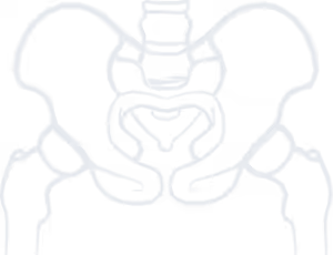
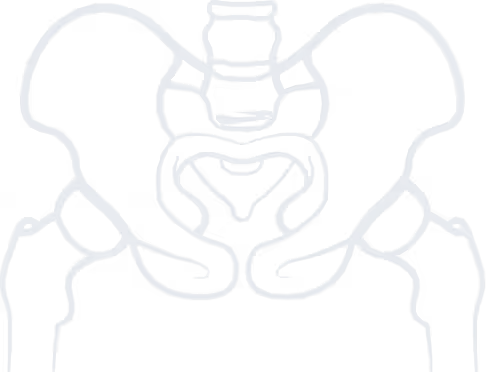

Avascular necrosis (AVN) or osteonecrosis of the femoral head is a serious condition that can rapidly lead to the destruction of the subchondral bone of the femoral head and the overlying cartilage, ultimately resulting in secondary hip osteoarthritis, potentially at a young age. Core decompression of the femoral head has been described as a potentially joint-preserving surgical intervention for patients with osteonecrosis.
The rationale behind this procedure is that, by drilling through the necrotic zone, the surrounding healthy tissue is stimulated to produce new bone. The drill channels serve as the necessary trigger for initiating a healing process from viable osteocytes in the adjacent bone, which subsequently migrate into the necrotic area and gradually replace the dead bone with healthy bone. Additionally, the addition of live osteoprogenitor cells harvested from the patient’s own bone marrow during the operation and injected into the necrotic site through the drill channels further enhances the regenerative response. Since the formation of new bone can only replace the volume of the necrotic region but cannot restore the spherical shape of the femoral head, core decompression is effective only in the early stages of the disease, before femoral head collapse occurs.
As with any surgical intervention, preoperative evaluation is essential to ensure patient safety during the procedure. An additional critical parameter before femoral head decompression is the three-dimensional assessment of the necrotic area to accurately plan the trajectory of the drill channels. This is achieved through detailed evaluation of radiographs, CT scans, and MRI studies by the surgeon.
In most cases, general anesthesia is used, and patients are placed in the supine position on a traction table. This is a specialized surgical table with a padded perineal post positioned between the patient’s legs, and boots applied to one or both lower limbs, allowing controlled application of traction. During the procedure, fluoroscopy is used both for intraoperative imaging of the hip in various positions and for guiding the drill trajectory to accurately target the osteonecrotic zone. Depending on the technique, either a small incision is made down to the bone, or three to four small stab incisions are created on the lateral surface of the thigh at the desired level. Under fluoroscopic guidance, guidewires are passed through the lateral femoral cortex, aiming at the necrotic area. Several technical variations have been described regarding the number, diameter, and positioning of the drill holes: some authors describe using a centrally placed reamer of progressively increasing diameter up to a large size, while others prefer using 2–3 smaller-diameter drill holes, depending on the size of the necrotic lesion. In all cases, each channel is drilled 2–3 times to ensure adequate stimulation of the healing response. As mentioned earlier, a bone marrow aspirate can be obtained before drilling. After centrifugation and sometimes combined with demineralized bone matrix, it can be injected into the femoral head. Some surgeons may also inject calcium phosphate, a bone substitute, to seal the drill channels and prevent marrow leakage. Finally, the wound is closed in layers.

Stop living in pain, improve the quality of your life!
As with any surgical procedure, hip core decompression carries the risk of complications. The incidence of surgical site infection is generally low (approximately 1%) due to the minimally invasive nature of the procedure, especially with percutaneous techniques, and the use of perioperative prophylactic antibiotics. Thromboembolic prophylaxis (to prevent deep vein thrombosis and pulmonary embolism) is routinely administered perioperatively with low-molecular-weight heparin. Iatrogenic femoral neck or subtrochanteric fractures are rare but have been reported. Preventive measures include using the minimum number of drill channels required to cover the necrotic zone and avoiding drilling distal to the level of the lesser trochanter.
Postoperatively, patients are mobilized with minimal weight-bearing for 4–6 weeks, followed by partial weight-bearing for another 3–4 weeks, progressing to full weight-bearing after 2 months. Immediate postoperative physiotherapy guidance and supervision by an experienced therapist is necessary. The bone regeneration process triggered by drilling can be radiographically assessed starting 6 weeks postoperatively and at 6–8 week intervals until 6 months after surgery. The success rates of core decompression vary considerably among patients with osteonecrosis and depend strongly on the stage of the disease (i.e., whether the subchondral bone and the shape of the femoral head have been affected), as well as on the size and location of the necrotic area. Patients in pre-collapse stages, with centrally located (rather than laterally located) necrosis and a combined necrotic angle of less than 240°, may achieve healing rates of nearly 90% after decompression.
Beyond the use of autologous bone marrow aspirate, which is readily available from the patient’s pelvis during surgery, other biological adjuncts have been used to enhance the regenerative response, including autografts, allografts, bone morphogenetic proteins (BMPs), and even stem cells. While their efficacy has been reported in small patient series, larger prospective randomized studies are needed for confirmation. Additionally, robot-assisted drilling techniques have also been described as alternatives to conventional fluoroscopy-guided methods.
Femoral head core decompression is a joint-preserving procedure indicated in the early stages of avascular necrosis. It works by stimulating new bone formation within the necrotic zone through drilling. It is a relatively safe and straightforward procedure performed under general anesthesia, associated with low complication rates. Postoperative rehabilitation is generally prolonged due to the already impaired functional status of patients with AVN at the time of surgery. However, when performed in appropriately selected patients, decompression can lead to significantly improved functional outcomes and may delay or even prevent the need for hip arthroplasty, particularly in younger individuals.
to guide you about your condition, so you can choose the best possible treatment for it.
We use cookies to improve your experience on our site. By using our site, you consent to cookies.
Manage your cookie preferences below:
Essential cookies enable basic functions and are necessary for the proper function of the website.
These cookies are needed for adding comments on this website.
Google reCAPTCHA helps protect websites from spam and abuse by verifying user interactions through challenges.
Google Tag Manager simplifies the management of marketing tags on your website without code changes.
Marketing cookies are used to follow visitors to websites. The intention is to show ads that are relevant and engaging to the individual user.
Google Maps is a web mapping service providing satellite imagery, real-time navigation, and location-based information.
Service URL: policies.google.com (opens in a new window)