


Hip fractures represent a distinct category of fractures, as they are associated with significant impairment of patients’ quality of life and increased mortality, especially in those with multiple comorbidities. It is estimated that approximately 30% of patients with a hip fracture will die within the first year following the injury. Moreover, only 20% of patients can walk independently and without assistance (walker or crutches) one year after the fracture. Finally, hip fractures are among the most costly injuries for patients, families, healthcare systems, and insurance providers, as patients lose their independence for extended periods and usually require structured rehabilitation programs. They occur most commonly in the elderly, in women, in Caucasians, in osteoporotic individuals, and in patients prone to frequent falls for various reasons.
Hip fractures occur in the proximal femur and include femoral head fractures, femoral neck fractures, and peritrochanteric fractures. Femoral head fractures are rare and usually associated with traumatic hip dislocations. Femoral neck fractures (or subcapital fractures) are relatively common, especially in older patients, and may occur even after low-energy trauma (e.g., a fall from standing or sitting height). Peritrochanteric fractures are subdivided into three categories depending on the fracture line relative to the line connecting the greater and lesser trochanters (the intertrochanteric line). If the fracture occurs above the line, it is termed a basocervical fracture. If the fracture follows the line, it is called an intertrochanteric fracture. Finally, if the fracture occurs below the line, it is classified as a subtrochanteric fracture. Peritrochanteric fractures also occur mostly in the elderly, usually after low-energy falls.
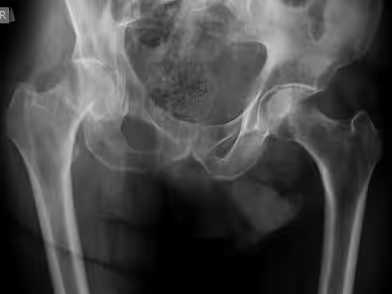
The main symptoms of a hip fracture are hip pain, inability to bear weight on the affected limb, and, in most cases, visible deformity. Typically, the injured leg appears shorter than the healthy one and externally rotated. Passive movement of the leg by an examiner causes severe pain in the groin. The patient is usually found lying on the floor, unable to rise. Rarely, patients may still manage to walk despite pain, in cases where the fracture fragments are impacted.
Diagnosis is based on clinical examination combined with the history of trauma and is confirmed with plain radiographs. If the fracture line is not clearly visible on X-rays, CT scanning provides detailed imaging of the bony structures.

Stop living in pain, improve the quality of your life!
The treatment of hip fractures is surgical. Conservative management is rarely chosen, only for patients who cannot undergo surgery due to severe comorbidities. Patients operated on within the first 48 hours of injury achieve better outcomes and fewer complications than those with delayed surgery.
The surgical method depends mainly on the type of fracture and the patient’s age.
a) Femoral head fracturesUsually treated with open reduction and internal fixation. The anterior hip approach is preferred; fracture fragments are reduced and stabilized with small screws.
β) b) Femoral neck fractures.
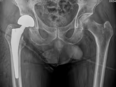
γ) c) Peri-trochanteric fractures • Regardless of age, the treatment of choice is closed reduction and fixation with intramedullary nailing. Under general or spinal anesthesia, the patient is positioned on a traction table, fracture reduction is achieved under fluoroscopy, and stabilized with a gamma intramedullary nail. The nail is inserted through the greater trochanter into the femoral canal, fixed with a screw traversing the femoral neck and head, and an additional distal locking screw. Rarely, a dynamic hip screw and plate system or even THA may be selected in highly comminuted fractures.
Prompt treatment and early initiation of rehabilitation after hip fracture are of utmost importance for favorable outcomes. Patients are often elderly with significant comorbidities. Early mobilization reduces the risk of serious complications such as thrombosis, pneumonia, or heart failure. Patients with peritrochanteric or subcapital fractures treated with screw fixation usually undergo a rehabilitation program lasting about six weeks. Those treated with hemiarthroplasty or THA typically recover faster, especially when surgery is performed using the ASI approach, in which case postoperative physiotherapy is often unnecessary.
All patients with hip fractures not caused by high-energy trauma should be evaluated for osteoporosis and assessed for fall risk factors to prevent future fractures.
to guide you about your condition, so you can choose the best possible treatment for it.
We use cookies to improve your experience on our site. By using our site, you consent to cookies.
Manage your cookie preferences below:
Essential cookies enable basic functions and are necessary for the proper function of the website.
These cookies are needed for adding comments on this website.
Google reCAPTCHA helps protect websites from spam and abuse by verifying user interactions through challenges.
Google Tag Manager simplifies the management of marketing tags on your website without code changes.
Marketing cookies are used to follow visitors to websites. The intention is to show ads that are relevant and engaging to the individual user.
Google Maps is a web mapping service providing satellite imagery, real-time navigation, and location-based information.
Service URL: policies.google.com (opens in a new window)