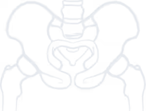
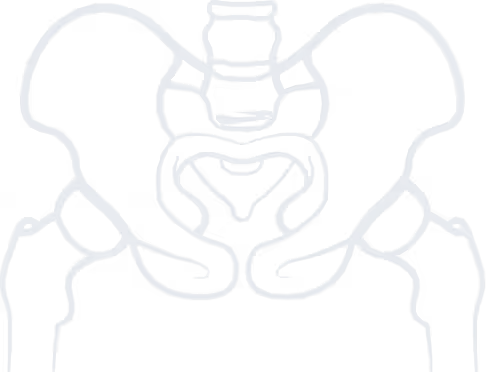

Osteonecrosis of the hip (also known as avascular necrosis of the femoral head) is the destruction of the femoral head due to impaired blood supply. It is a relatively common hip disorder (approximately 20,000 new cases are diagnosed annually in the United States), and about 10% of total hip arthroplasties are performed because of it. Osteonecrosis of the hip is more frequent in men and typically occurs between the ages of 35 and 50. In a significant proportion of patients (around 80%), it affects both hips, while in rare cases (about 3%), it may simultaneously involve other major joints (e.g., knees, shoulders).
The main underlying mechanism leading to osteonecrosis of the hip is disruption of the blood supply to the femoral head. This may occur after trauma or due to pathological causes.
Femoral head fractures carry a 75–100% risk of osteonecrosis. Fractures at the base of the femoral neck carry about a 50% risk, while hip dislocations are up to 40%. Intertrochanteric fractures rarely cause osteonecrosis. In these injuries, the likelihood of osteonecrosis increases with greater displacement of bone fragments and delayed treatment.
Pathological causes include hematologic diseases (e.g., hypercoagulability syndromes causing thrombosis, such as thrombophilia, hematologic malignancies such as leukemia and lymphoma, sickle cell anemia), alcoholism, decompression sickness (“diver’s disease”), and autoimmune conditions (e.g., systemic lupus erythematosus). Osteonecrosis can also occur in patients receiving corticosteroids, chemotherapy, immunosuppressive drugs, and is relatively common after radiotherapy. In some cases, it is idiopathic, meaning no underlying cause is identified.

Stop living in pain, improve the quality of your life!
Osteonecrosis of the hip typically presents with mild discomfort in the hip region, rather than severe symptoms. In the early stages, it may be completely asymptomatic. Typically, patients experience mild pain in the anterior groin, not necessarily related to intense activity. Without timely diagnosis, progressive destruction of the femoral head causes worsening pain—first during activity, later also at rest. Symptoms are aggravated when climbing stairs or walking on inclined surfaces. As in advanced hip osteoarthritis, late-stage osteonecrosis presents with severe joint pain, leg shortening, and stiffness (especially reduced internal rotation when the hip is flexed). At this stage, patients struggle with simple daily tasks such as putting on socks and shoes or trimming toenails.
Early diagnosis is crucial for favorable outcomes. Clinical examination and detailed history-taking may reveal risk factors (e.g., certain medications), though early disease often lacks abnormal findings on hip clinical tests.
Plain radiographs (anteroposterior pelvis/hips and lateral of the affected hip) are the first-line imaging, but may be normal in early stages. In later stages, radiographs may show focal sclerosis, subchondral collapse, loss of femoral head sphericity, and arthritic changes (joint space narrowing, osteophytes, cysts in the femoral head and/or acetabulum).
MRI is the gold standard for diagnosing osteonecrosis, particularly in early stages when plain X-rays are normal. Modern staging systems rely on MRI findings. Early MRI reveals impaired blood supply, as evidenced by focal bone marrow edema, and a clear demarcation of the necrotic area. Later stages show the destructive consequences of necrosis (subchondral fracture, collapse of the femoral head, and secondary arthritis).
Management depends primarily on the stage at diagnosis and the patient’s age, while also considering etiology (reversible or not) and activity level. The main goal is preservation of the femoral head, if possible, and restoration of joint function.
At Early-stage (Conservative treatment) With:
In young patients with early-stage disease and reversible causes, surgical treatment may also be considered alongside conservative management:
Advanced-stage (Femoral Head Collapse), once collapse has occurred, treatment is surgical. Options include:
to guide you about your condition, so you can choose the best possible treatment for it.
We use cookies to improve your experience on our site. By using our site, you consent to cookies.
Manage your cookie preferences below:
Essential cookies enable basic functions and are necessary for the proper function of the website.
These cookies are needed for adding comments on this website.
Google reCAPTCHA helps protect websites from spam and abuse by verifying user interactions through challenges.
Google Tag Manager simplifies the management of marketing tags on your website without code changes.
Marketing cookies are used to follow visitors to websites. The intention is to show ads that are relevant and engaging to the individual user.
Google Maps is a web mapping service providing satellite imagery, real-time navigation, and location-based information.
Service URL: policies.google.com (opens in a new window)