
Orthopedic condition
Calcific Tendonitis
Calcific tendonitis of the shoulder is a condition in which calcium deposits form within the tendons of the rotator cuff, causing – often – severe pain and restricted movement. Despite the discomfort it causes, it is a condition with a generally good prognosis, which in the great majority of cases is treated by conservative means.
Dr. Ioannis Tsarouhas, Orthopedic Surgeon with experience in conditions of the shoulder, treats calcific tendonitis on an individual basis, with the aim of relieving pain and restoring movement.
What is calcific tendonitis?
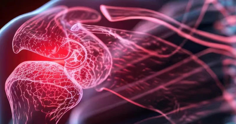
The shoulder is moved and stabilised by the rotator cuff – a complex of four tendons that surround the head of the humerus. In calcific tendonitis, calcium crystals are deposited within one or more of these tendons (most often the supraspinatus), forming a “calcific deposit”.
The exact cause of the deposition is not fully known. The calcific deposit can remain “silent” for a long time, but when it becomes inflamed or when the body begins to “resorb” it, it can cause severe pain. Characteristically, the condition goes through different phases: a formative phase, a resting period and a resorptive phase, which is often also the most painful.
Who is affected
Calcific tendonitis occurs most often in adults aged 30 to 60 and is somewhat more common in women. It may be associated with certain metabolic disorders, although it often occurs without any clear predisposing factor. In a proportion of patients, it can affect both shoulders.
Clinical presentation – Symptoms
The symptoms vary considerably depending on the phase of the disease:
- Severe pain, which in the acute phase (resorptive phase) can be very intense, even unbearable, often coming on suddenly.
- Pain that gets worse with movement of the shoulder, particularly when lifting the arm.
- Night pain, which disturbs sleep.
- Restricted movement and stiffness.
In other phases, the pain may be milder and chronic, or even absent, with the calcific deposit being discovered incidentally on an X-ray.
The phases of the disease in detail
Understanding the phases of calcific tendonitis helps to explain its particular behaviour. The disease is not static – it progresses through distinct phases:
- Formative phase: Calcium begins to be deposited within the tendon. In this phase, symptoms may be mild or even absent.
- Resting phase: The calcific deposit has formed and remains stable. It may cause mild, chronic pain or be completely asymptomatic.
- Resorptive phase: The body begins to “resorb” the calcific deposit. Paradoxically, this phase – which essentially marks the resolution of the disease – is also the most painful, as it is accompanied by intense inflammation. The pain can be sudden and very severe.
This natural course explains why the condition tends to be self-limiting: the calcific deposit is eventually resorbed. Knowing which phase the patient is in helps the doctor to choose the appropriate treatment.
Shock wave therapy (ESWT)
One of the effective non-invasive methods for calcific tendonitis is shock wave therapy (Extracorporeal Shock Wave Therapy). This is an extracorporeal method, in which energy waves are directed at the area of the calcific deposit.
The mechanism of action is twofold: the shock waves “break up” the calcific deposit, facilitating its resorption, while at the same time stimulating the local blood supply and the healing processes. The method is non-invasive, is applied in sessions and can offer significant relief in selected patients, making it a useful option between medication and the more invasive methods.
Diagnosis
The diagnosis is as a rule easy. X-rays clearly show the calcific deposit, its location and its size. Ultrasound is also very useful, as it locates the calcific deposit and can also be used therapeutically (guiding certain interventions). Magnetic resonance imaging (MRI) is used in selected cases, to assess the condition of the tendons.
Treatment
The great majority of cases of calcific tendonitis are treated successfully by conservative means, as the condition tends to resolve on its own over time, as the calcific deposit is resorbed.
Conservative treatment
This includes anti-inflammatory medication and painkillers for pain control, physiotherapy to maintain range of motion and – in more severe cases – local injections. A particularly effective intervention is needling and lavage of the calcific deposit under ultrasound guidance (barbotage), in which the calcific deposit is “broken up” and removed. Shock waves (ESWT), which stimulate the resorption of the calcific deposit, are also used.
Surgical treatment
In rare, resistant cases that do not respond to conservative treatment, the calcific deposit can be removed arthroscopically. This is a minimally invasive procedure with very good results.
Prognosis
The prognosis of calcific tendonitis is as a rule excellent. Although the acute phase can be particularly painful, the condition tends to resolve, often spontaneously, as the calcific deposit is resorbed by the body. With proper conservative treatment, most patients obtain complete relief, with no lasting effects on the function of the shoulder.
Cost of treatment
The cost depends on the type of treatment (medication, physiotherapy, shock waves, barbotage or – more rarely – arthroscopic removal), the number of sessions or interventions, and insurance cover. Precise information is given on an individual basis, after the clinical assessment. The clinic is at your disposal for any relevant information.
More about the factors that determine the cost and about cover by insurance funds: Cost of surgery.
When to see a doctor
Severe, sudden shoulder pain – particularly when it appears without a previous injury and severely restricts movement – should be assessed by an Orthopedic Surgeon. Although calcific tendonitis has a good prognosis, the correct diagnosis is important, both to confirm the condition and to rule out other causes of shoulder pain.
Early assessment allows appropriate treatment – particularly in the painful phase – so that the patient is relieved more quickly, without unnecessary suffering.
Frequently asked questions
Why is calcific tendonitis so painful?
The most severe pain usually occurs in the phase in which the body “resorbs” the calcific deposit, which is accompanied by intense inflammation. This phase, although painful, is also a sign that the calcific deposit is resolving.
Will it go away on its own?
In the great majority of cases, yes. The condition tends to be self-limiting as the calcific deposit is resorbed. Conservative treatment helps to control the pain and speeds up resolution.
What is barbotage?
It is an ultrasound-guided intervention in which the calcific deposit is needled and washed out, so that it is removed. It is effective in selected cases.
Is surgery often needed?
No. Surgical (arthroscopic) removal is reserved for the rare, resistant cases that do not respond to conservative treatment.
Is the cost covered by insurance?
Cover depends on the type of treatment and on the insurance policy. The clinic can give you detailed information.
How long does calcific tendonitis last?
The duration varies considerably. The disease progresses through phases and, eventually, the calcific deposit is resorbed, often spontaneously. The acute, painful phase lasts as a rule for weeks, while the overall course may be longer.
Does it come back after treatment?
In the great majority of cases, once the calcific deposit has been resorbed or removed, the condition does not recur. In some patients, however, a calcific deposit may appear at another site or in the other shoulder.
Contact
Book an appointment with Dr. Tsarouhas
Fill in the form with the time that suits you and we will contact you, or call us directly.
Call us 210 6464 744-
Petroupoli 1 Agias Triados St., 132 31 210 5011 717
-
Heraklion, Crete 49 Kyrillou Loukareos St., 712 01
Appointment request
We reply to every request on the same or the next working day.
Your details are used solely to contact you.