
Orthopedic condition
Osteonecrosis of the Knee
Osteonecrosis of the knee is a condition in which the blood supply to a segment of bone is interrupted, resulting in its gradual death. Without timely diagnosis and treatment, it can lead to collapse of the joint surface and early osteoarthritis of the knee.
Dr. Ioannis Tsarouhas, Orthopedic Surgeon specialising in conditions of the major joints, treats osteonecrosis of the knee across the full range of its stages, from conservative management to modern surgical techniques.
What is osteonecrosis of the knee?
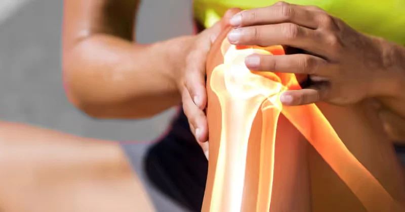
The term “osteonecrosis” (or aseptic necrosis) means the death of bone tissue due to interruption of its blood supply. Bone, like every living tissue, needs a continuous supply of blood to stay healthy. When this supply is interrupted, the bone cells die and the bone progressively weakens.
In the knee, osteonecrosis usually affects the lower end of the thigh bone (the femoral condyles) and, more rarely, the upper end of the tibia. As the necrotic bone weakens, the overlying joint surface – together with the cartilage – is at risk of “collapsing”, leading to irregularity of the joint and, ultimately, to arthritis.
Types of osteonecrosis of the knee
Osteonecrosis of the knee is divided into main categories, depending on the cause:
- Primary (spontaneous) osteonecrosis: It occurs mainly at older ages, without a clear predisposing factor, and usually affects only one part of the knee.
- Secondary osteonecrosis: It is associated with specific risk factors and often occurs in younger patients, sometimes at more than one site.
- Postoperative osteonecrosis: It may occur after certain operations on the knee.
This distinction matters, as it affects the prognosis and the treatment approach.
Causes and risk factors
While primary osteonecrosis occurs without a clear cause, secondary osteonecrosis is associated with certain risk factors, such as:
- Long-term use of corticosteroids
- Excessive alcohol consumption
- Certain blood diseases and metabolic disorders
- Injuries to the area
- Certain systemic conditions
It is important to stress that the presence of a risk factor does not mean that someone will develop osteonecrosis, while in many cases no clear cause is identified.
How osteonecrosis progresses
Understanding how osteonecrosis progresses explains why time is so critical. The process begins with the interruption of the blood supply to a segment of the bone. At this phase, the bone begins to die, but the joint surface still remains intact – symptoms may be mild or even absent.
As the necrotic bone weakens, it loses its ability to withstand loads. At this critical point, the overlying joint surface is at risk of “collapsing” – that is, of giving way and forming an irregularity. Once this collapse occurs, the damage becomes irreversible: the smooth joint surface is permanently lost and a rapid course towards osteoarthritis begins.
It is precisely this sequence – necrosis, weakening, collapse, arthritis – that makes early diagnosis so valuable: the earlier we intervene before the collapse, the more options there are for preserving the joint.
Clinical picture – Symptoms
The main symptom of osteonecrosis of the knee is pain, which has certain characteristics:
- Sudden onset of pain, often without a previous injury, especially in the primary form.
- Pain that is localised, usually on the inner side of the knee.
- Worsening with weight-bearing and activity.
- Night pain, which may disturb sleep.
- Swelling and restricted range of motion, in more advanced stages.
The intensity of the symptoms and their progression vary depending on the extent of the necrosis and the stage of the disease.
Diagnosis and stages
Early diagnosis of osteonecrosis is decisive for the prognosis. It begins with the medical history and clinical examination and continues with imaging.
X-rays may be normal in the early stages, which is why magnetic resonance imaging (MRI) is the examination of choice: it reveals the necrosis very early, before radiographic changes appear, and assesses the extent of the lesion.
The disease is staged according to its progression, from the early stages (necrosis without collapse of the surface) to the advanced ones (collapse of the joint surface and development of arthritis). The stage largely determines the treatment approach: the earlier the diagnosis, the more options there are for preserving the joint.
Before treatment: the importance of a proper assessment
Before any treatment decision, a comprehensive assessment of the lesion is essential. The surgeon takes into account the stage of the disease, the extent and location of the necrosis, the condition of the joint surface (whether or not it has begun to collapse), the patient’s age and activity level, as well as any underlying risk factors.
Addressing the risk factors – where they exist – is just as important as the local treatment itself. For example, reviewing a course of corticosteroid medication or treating an underlying condition can affect the course of the disease. This holistic approach – which addresses both the lesion and its causes – is characteristic of the proper management of osteonecrosis.
Treatment
The treatment of osteonecrosis is individualised according to the stage, the extent of the lesion, and the patient’s age and general condition.
Conservative treatment
In the early stages, especially for small lesions, treatment may be conservative: offloading the limb with walking aids, activity modification, medication to control pain, and physiotherapy. Some small, early lesions may resolve with conservative treatment.
Surgical treatment
When the lesion is extensive or advanced, or does not respond to conservative treatment, surgical treatment is indicated. The options depend on the stage:
- Joint-preserving techniques in the earlier stages, such as drilling (decompression) of the necrotic area, which aims to restore the blood supply and relieve pain.
- Unicompartmental or total knee replacement in the advanced stages, when the joint surface has collapsed and arthritis has developed.
The appropriate method is chosen on an individual basis, with the aim – wherever feasible – of preserving the natural joint, especially in younger patients.
Why early diagnosis matters
Osteonecrosis of the knee is a condition in which time is of decisive importance. In the early stages, before the joint surface collapses, joint-preserving techniques can relieve the patient and prevent progression to arthritis. Once the surface collapses, however, the damage becomes irreversible and the only remaining solution is joint replacement.
For this reason, sudden, unexplained knee pain – especially when accompanied by worsening at night – should not be ignored, but investigated promptly with an MRI scan.
Osteonecrosis and osteoarthritis: what is the difference?
Osteonecrosis is often confused with osteoarthritis, as both cause knee pain and can ultimately lead to wear of the joint. However, they are two different conditions with different starting points.
In osteoarthritis, the problem starts with the wear of the articular cartilage, that is, from the “surface” of the joint inwards. In osteonecrosis, by contrast, the problem starts in the bone itself, beneath the cartilage, due to the interruption of the blood supply – that is, “from the inside out”. This distinction is not theoretical: osteonecrosis, especially in the early stages, can be treated with techniques that aim to restore the bone (such as drilling), which is not the case for osteoarthritis. This is why the correct diagnosis – mainly with MRI – is decisive for the right treatment decision.
Follow-up and prognosis
The prognosis of osteonecrosis of the knee depends directly on the stage at which it is diagnosed and on the extent of the lesion. Small lesions that are detected early have a good prognosis and may be treated with preservation of the joint. In contrast, extensive lesions or advanced stages with collapse of the surface have a less favourable prognosis and often lead to joint replacement.
For this reason, regular follow-up – with clinical reassessment and imaging – is important, so that the progression of the lesion is monitored and the appropriate treatment decisions are made in good time.
Cost of treating osteonecrosis of the knee
The cost depends on the stage and the type of treatment (conservative, drilling, joint replacement), the materials that may be used, the hospital and the room category, as well as the patient’s insurance cover. In most cases, private insurance policies and insurance funds cover a significant part of the expense.
Precise information is provided on an individual basis, after the clinical assessment. The clinic is at your disposal for any relevant information.
More on the factors that determine the cost and on cover by insurance funds: Cost of surgery.
The role of rehabilitation
Rehabilitation after treatment of osteonecrosis depends on the type of treatment. After joint-preserving techniques, such as drilling, weight-bearing is initially protected, to give the bone time to recover. After joint replacement, rehabilitation follows the corresponding protocol.
In every case, the physiotherapy programme focuses on maintaining range of motion, muscle strengthening and the gradual return to walking. Adherence to the programme and close cooperation with the surgeon are decisive for the final outcome.

Frequently asked questions
What causes osteonecrosis of the knee?
It is due to interruption of the blood supply to a segment of the bone. It may be primary (without a clear cause, usually at older ages) or secondary (associated with factors such as corticosteroid use or excessive alcohol consumption).
What is the main symptom?
Pain, often of sudden onset, located on the inner side of the knee, which worsens with weight-bearing and may worsen at night.
How is it diagnosed?
MRI is the examination of choice, as it reveals the necrosis very early, when X-rays are still normal.
Is surgery always needed?
No. In the early stages, small lesions may be treated conservatively. Surgery is indicated for extensive or advanced lesions.
Can joint replacement be avoided?
In the early stages, joint-preserving techniques (such as drilling) can prevent or delay progression. Once the joint surface collapses, joint replacement becomes the indicated solution.
Is the cost covered by insurance?
In most cases, private insurance policies and insurance funds cover part or all of the expense. The clinic can give you detailed information.
Is osteonecrosis the same as arthritis?
No. In arthritis the problem starts with the wear of the cartilage, whereas in osteonecrosis it starts with the interruption of the blood supply to the bone beneath the cartilage. The distinction is important, as it affects treatment.
Can it affect both knees?
Yes, especially in the secondary form, which is associated with systemic risk factors, osteonecrosis can occur at more than one site or joint.
How important is the timing of diagnosis?
Decisive. In the early stages, before the joint surface collapses, joint-preserving techniques can change the course of the disease. After the collapse, the damage is irreversible.
What are the main risk factors?
Long-term corticosteroid use and excessive alcohol consumption are among the most common. In many cases, however, no clear cause is identified (primary form).
Contact
Book an appointment with Dr. Tsarouhas
Fill in the form with the time that suits you and we will contact you, or call us directly.
Call us 210 6464 744-
Petroupoli 1 Agias Triados St., 132 31 210 5011 717
-
Heraklion, Crete 49 Kyrillou Loukareos St., 712 01
Appointment request
We reply to every request on the same or the next working day.
Your details are used solely to contact you.