
Orthopedic condition
Chondromalacia Patellae
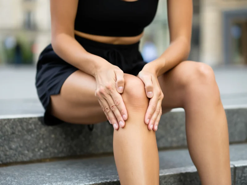
Chondromalacia patellae is wear of the articular cartilage on the back surface of the kneecap (patella), where it glides over the femoral trochlea. It is the most common cause of pain at the front of the knee and occurs both in young people who exercise and in middle-aged adults.
Why it occurs
The patella is subjected to enormous loads: in a deep squat, the forces acting on the patellofemoral joint reach several times body weight. When the patella does not glide centrally within the groove, the pressure is concentrated on one area of the cartilage and wears it down. The main causes are weakness of the vastus medialis and of the hip abductors, tightness of the iliotibial band and of the hamstrings, an increased Q angle, trochlear dysplasia, a high or low position of the patella, excessive rotational deformities of the femur, overuse, and previous injury or dislocation.
Symptoms
A dull pain around or behind the patella, which worsens when going down stairs, in deep squats, when running downhill and after prolonged sitting with the knees bent — the so-called “cinema sign”. There is often grating or clicking on bending, a feeling that the knee will “give way” because of quadriceps inhibition, and mild diffuse swelling after exercise.
Stages
The lesion is staged according to the Outerbridge classification: stage I with softening of the cartilage, stage II with superficial fissures of less than half the thickness, stage III with deep fissures that reach close to the subchondral bone, and stage IV with complete exposure of the bone. The early stages are entirely reversible in terms of symptoms, even though cartilage does not regenerate.
Diagnosis
The clinical examination includes palpation of the margins of the patella, the compression test, assessment of patellar tracking, and testing of strength and flexibility. The axial skyline view and weight-bearing X-rays show the alignment and the patellofemoral joint space. MRI depicts the extent and depth of the cartilage lesion and the oedema of the subchondral bone.
Treatment
Conservative treatment is the first-line treatment and is effective in the great majority of patients. It includes targeted strengthening of the quadriceps and gluteal muscles, stretching, correction of exercise technique, weight reduction, temporary avoidance of aggravating movements, anti-inflammatory medication and, selectively, intra-articular injections of hyaluronic acid or PRP. Improvement takes weeks to months of consistency.
Surgery is reserved for persistent, focal lesions that do not respond. The options include arthroscopic smoothing or microfracture, modern cartilage repair techniques, lateral patellar retinacular release or reconstruction of the medial patellofemoral ligament in cases of instability, and tibial tubercle transfer in cases of significant maltracking. See also knee cartilage lesions.
Contact
Book an appointment with Dr. Tsarouhas
Fill in the form with the time that suits you and we will contact you, or call us directly.
Call us 210 6464 744-
Petroupoli 1 Agias Triados St., 132 31 210 5011 717
-
Heraklion, Crete 49 Kyrillou Loukareos St., 712 01
Appointment request
We reply to every request on the same or the next working day.
Your details are used solely to contact you.