
Orthopedic condition
Knee Tendon Rupture (Quadriceps & Patellar)
The extensor mechanism of the knee consists of the quadriceps muscle, the quadriceps tendon, the patella (kneecap) and the patellar tendon. A rupture of any of these elements abolishes the ability to actively extend the knee and is an orthopedic emergency.
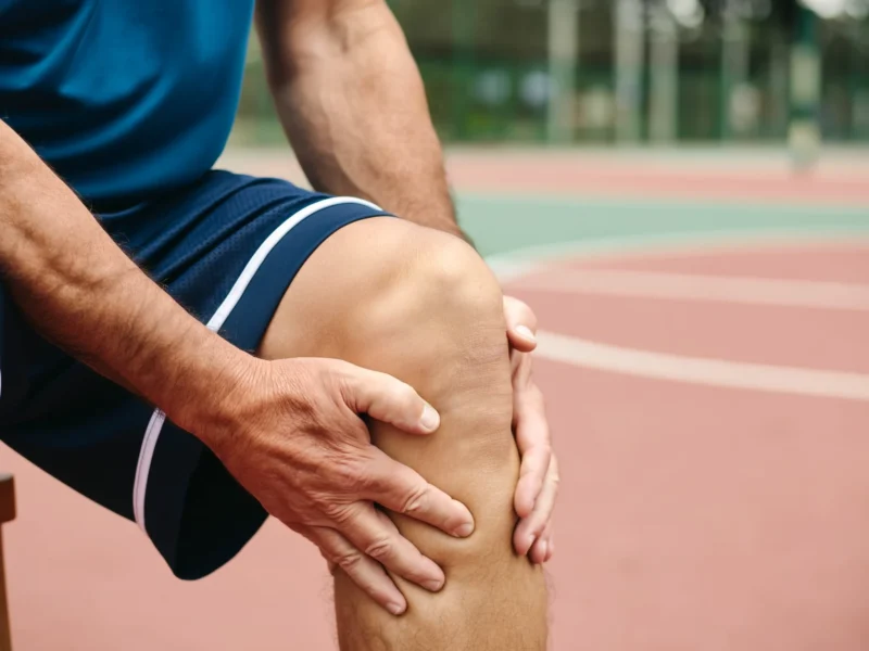
Quadriceps tendon rupture
It usually affects patients over the age of 40 and occurs during a sudden eccentric contraction, typically when the knee bends suddenly while the quadriceps is contracting – for example on a step or when stumbling. The tendon usually tears just above the patella. Predisposing factors are diabetes mellitus, chronic renal failure, hyperuricaemia, long-term corticosteroid use, quinolones and pre-existing tendinopathy.
Patellar tendon rupture
It occurs more often in younger, athletic adults under 40, after a jump or a violent contraction. The tendon generally tears at the lower pole of the patella. It is often preceded by a period of patellar tendinopathy, the so-called “jumper’s knee”.
Symptoms
Acute pain at the front of the knee at the moment of injury, often with a tearing sensation, immediate swelling and haemarthrosis. The patient cannot actively straighten the knee or keep the straightened leg raised, and the knee gives way on weight-bearing. A gap can be felt above or below the patella. Characteristically, the patella is displaced downwards in a quadriceps tendon rupture and upwards in a patellar tendon rupture.
Diagnosis
The inability to actively extend the knee is the key finding and is often sufficient for the diagnosis. The lateral X-ray shows the displacement of the patella and rules out a fracture. Ultrasound and MRI confirm the location and extent of the rupture and distinguish a complete rupture from a partial one, which is treated conservatively.
Why surgery must be performed promptly
A complete rupture requires surgical repair. A delay beyond two to three weeks leads to shortening and fibrosis of the tendon and the quadriceps, makes reapproximation more difficult, may require a graft and is associated with a worse functional outcome. The repair is performed with transosseous sutures or with anchors, and the extensor mechanism is protected postoperatively.
Rehabilitation
This is followed by a period of protection with a brace in extension and controlled, gradually increasing flexion under physiotherapy guidance for the first six weeks. Weight-bearing is allowed early with the brace in extension. Quadriceps strengthening begins progressively from the second to third week. Full range of motion is usually regained at three months and return to sporting activities at six to nine months, based on strength rather than time.
Contact
Book an appointment with Dr. Tsarouhas
Fill in the form with the time that suits you and we will contact you, or call us directly.
Call us 210 6464 744-
Petroupoli 1 Agias Triados St., 132 31 210 5011 717
-
Heraklion, Crete 49 Kyrillou Loukareos St., 712 01
Appointment request
We reply to every request on the same or the next working day.
Your details are used solely to contact you.